Scientific Imagery: Six Modes of Reading
The scientific imagery of Christ Carrying the Cross brings together six complementary modes of reading. Each provides specific information on the structure of the panel, the paint layers, restorations or the material history of the work.
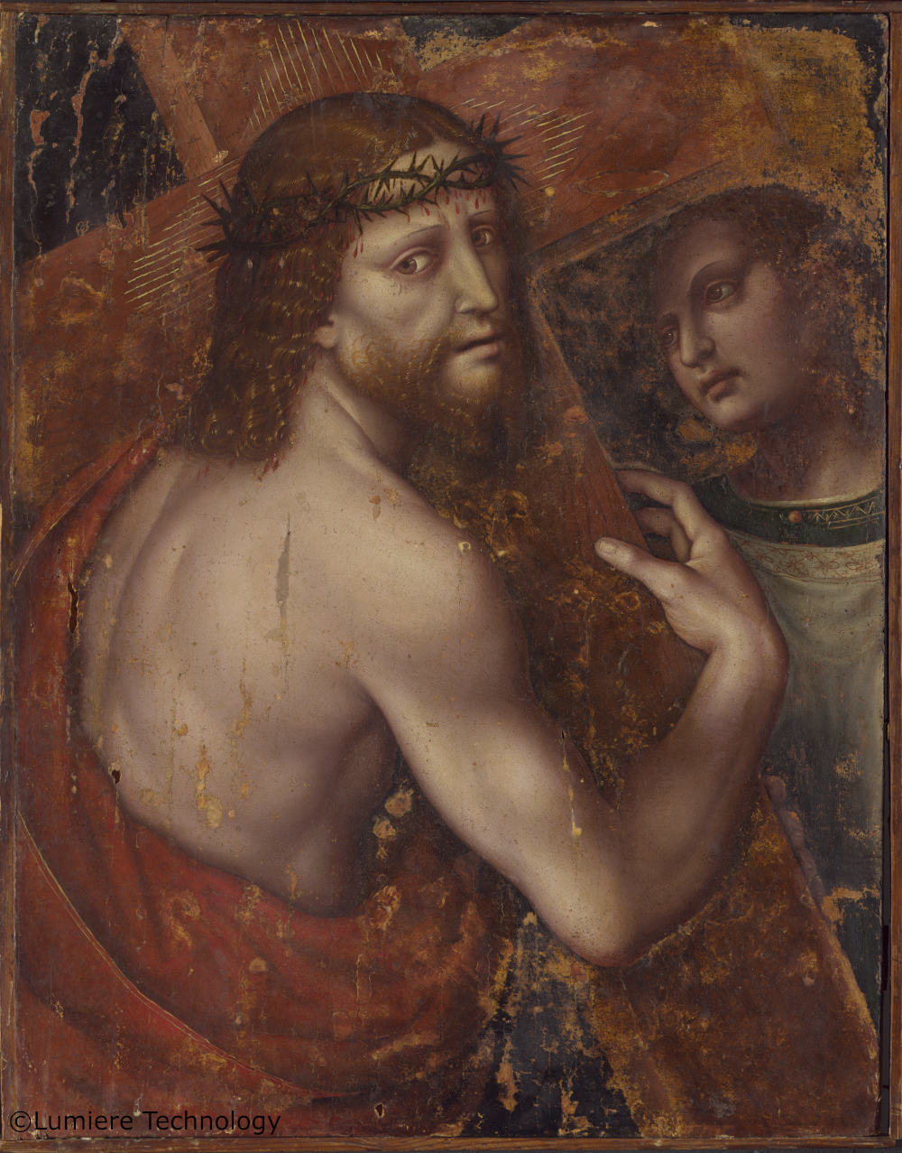
Visible Image (D65)
– Apparent state of the work: composition, colours, modelling
– Reference document for describing the painting
– Basis for comparison with the other imaging methods

X-ray Radiography
– Internal structure of the panel and paint layers
– Pigment densities and superimpositions
– Reveals elements invisible in visible light
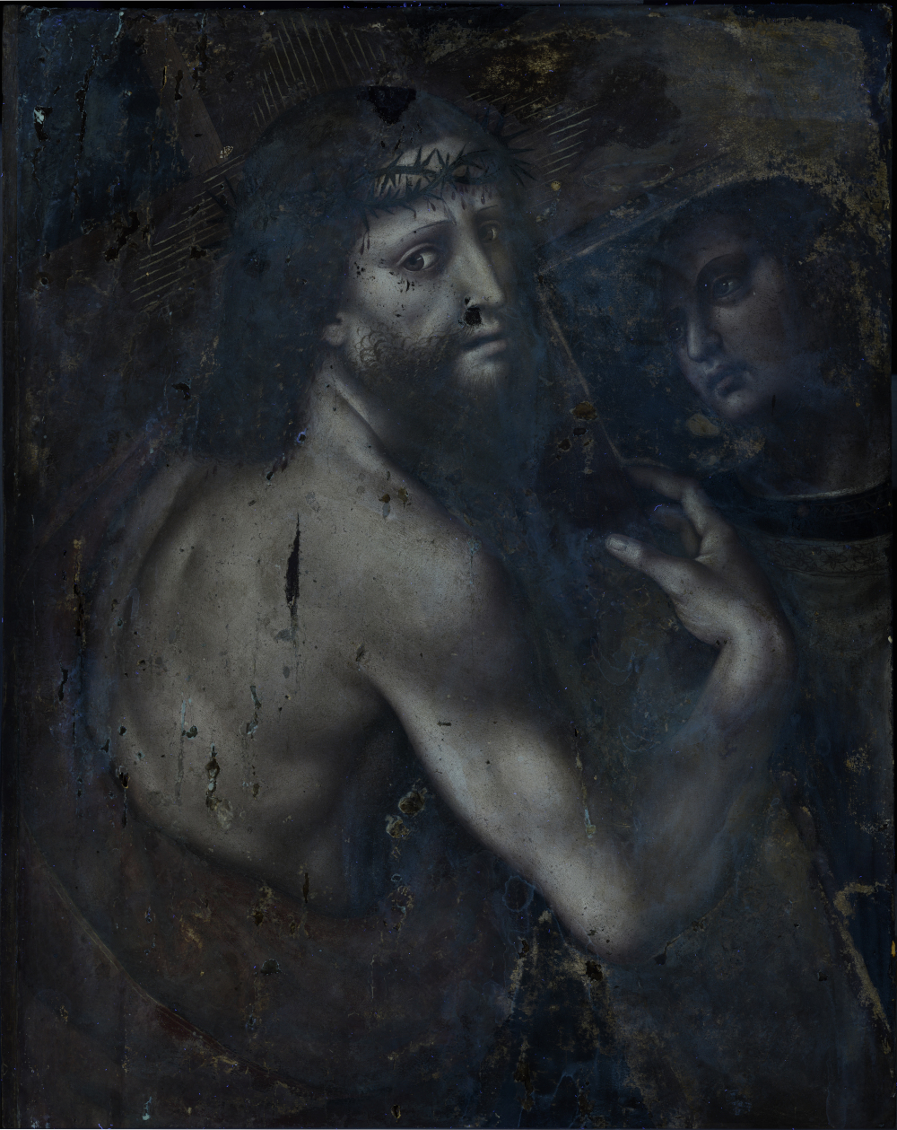
Ultraviolet (UV)
– Detection of varnishes and retouching
– Identification of later interventions
– Mapping of restored areas
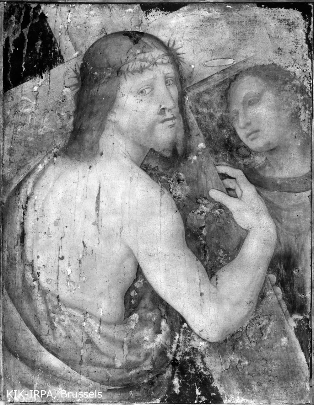
Infrared (IR 1700)
– Reveals the underlying drawing
– Reading of pentimenti and adjustments
– Distinguishes preparatory tracing from paint execution
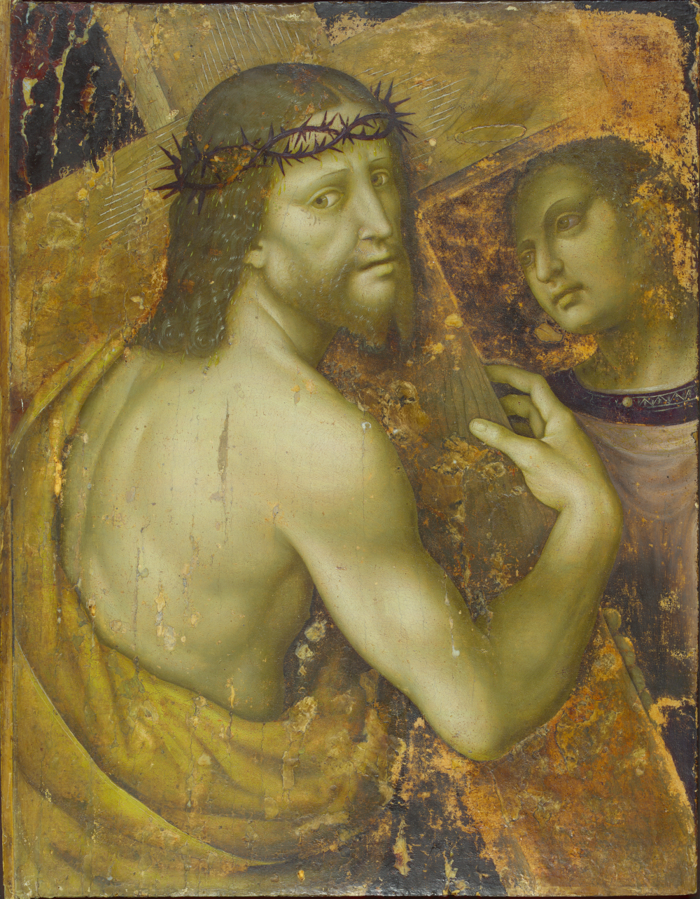
Infrared False Colour (IRFC)
– Translates information invisible to the eye into visible colours
– Identification of overpaints and retouching
– Helps with stratigraphic reading
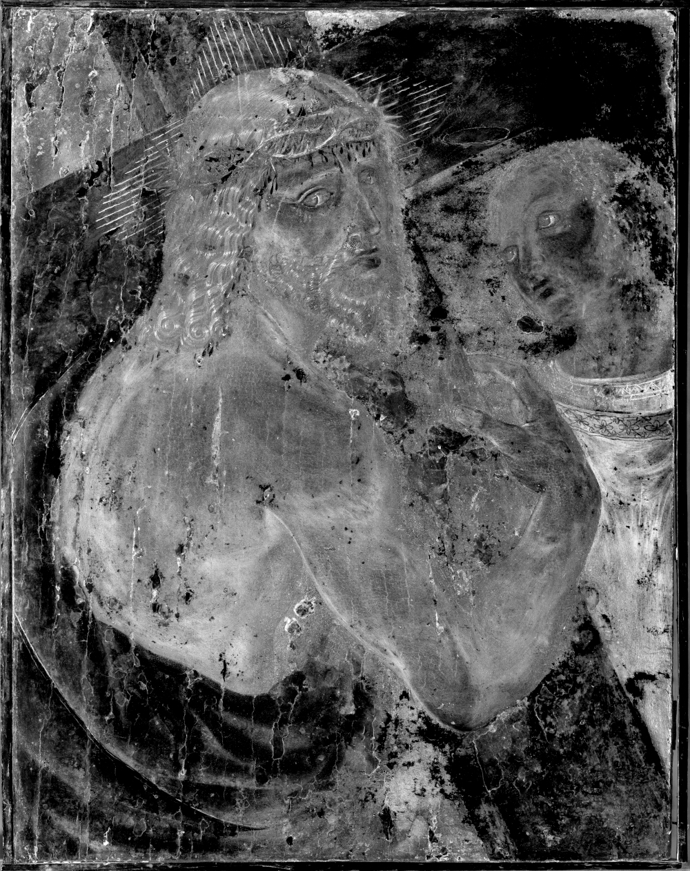
Emissiography
– Mainly analyses the painted surface
– Reveals certain pigment differences
– Complements X-ray radiography
Together, these six modes of imaging make it possible to distinguish the visible appearance of the work, its material structure, later interventions and certain underlying elements not perceptible to the naked eye.